Blank Diagram Of A Long Bone : A P I Long Bone Labeling Flashcards Quizlet - Long bones function as rigid bars that move when muscles contract.. The blood vessels inside a bone. This is an online quiz called label the long bone. The structure of a long bone allows for the best visualization of all of the parts of a bone (figure 1). The hollow region in the diaphysis is called the medullary cavity, which is filled with yellow. The bones shown in the chest and hip region in the labeled human skeleton diagram are the ribs, vertebrae, pelvis, os coxae, sacrum and coccyx.
The largest bone in the body, the _____, is a long bone. Bones of the axial and appendicular skeleton. The structure of a long bone allows for the best visualization of all of the parts of a bone (). A typical long bone shows the gross anatomical characteristics of bone. The diaphysis is the tubular shaft that runs between the proximal and distal ends of the bone.
This Image Depicts The Shoes Of Bones Examples Of Flat Bones Are The Scapulas And The Sternum Examples Of Irregular B Sphenoid Bone Medical Knowledge Scapula from i.pinimg.com Covers the surfaces of bones where they come together to form…. The osteons are made up of the living osteocytes and mineral matrix which supplies blood. Choose from 500 different sets of long bone diagram flashcards on quizlet. It is very strong to support the body's weight. The diaphysis and the epiphysis. The largest bone in the body, the _____, is a long bone. Short bones provide stability and support as well as. Diagramme schnell und einfach erstellen.
It is placed laterally to tibia and is the most slender of all the long bones.
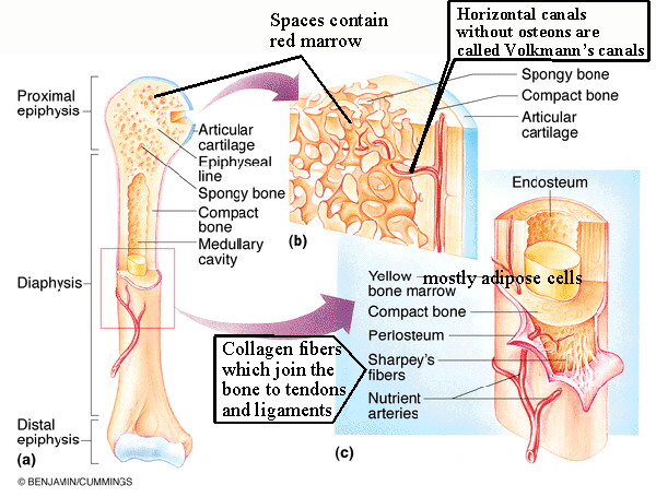
Layer of bone tissue that has many small spaces and is found j…. Create flashcards for free and quiz yourself with an interactive flipper. It also protects several vital organs of the chest, such as the heart, aorta, vena cava, and. Bones of the axial and appendicular skeleton. It is very strong to support the body's weight. It is placed laterally to tibia and is the most slender of all the long bones. Shaft of a long bone. Long bones include the humerus this image represents the parts of a long bone. The diaphysis is the tubular shaft that runs between the proximal and distal ends of the bone. End of a long bone. Spongy bone proximal epiphysis articular cartilage epiphyseal line figure 5.2a the structure of a long bone (humerus). There is a printable worksheet available for download here so you can take the quiz with pen and paper. The tarsus or heel bone consist of 7 bones that make up the posterior part of the foot, that is present between the tibia, fibula and metatarsals.
This is an online quiz called label the long bone. Layer of bone tissue that has many small spaces and is found j…. Bones of the axial and appendicular skeleton. The bundle gives you access to a second set of diagrams, studying the anatomy of a long bone. This is a quiz called label the long bone and was created by member deanne1480 advertisement.
Biol 237 Class Notes Skeletal System from www.unm.edu A long bone has two parts: A long bone is a bone that is significantly longer than it is wide. Several muscles that move the arms, head, and neck have their origins on the sternum. Short bones provide stability and support as well as. It is placed laterally to tibia and is the most slender of all the long bones. Covers the surfaces of bones where they come together to form…. Related posts of diagram of of a long bone bones and muscles anatomy. It also protects several vital organs of the chest, such as the heart, aorta, vena cava, and.
A long bone has two parts:
Related posts of diagram of of a long bone bones and muscles anatomy. This is an online quiz called label the long bone. You need to get 100% to score the 10 points available. Bones of the axial and appendicular skeleton. The diaphysis is the tubular shaft that runs between the proximal and distal ends of the bone. End of a long bone. The tarsus or heel bone consist of 7 bones that make up the posterior part of the foot, that is present between the tibia, fibula and metatarsals. The femur is a type of long bone located in the thigh and is the largest bone of the skeletal system. Most, but not all, features you are required to know are shown on the following pages. The diaphysis and the epiphysis. The sternum, commonly known as the breastbone, is a long, narrow flat bone that serves as the keystone of the rib cage and stabilizes the thoracic skeleton. A typical long bone shows the gross anatomical characteristics of bone. It contains the bone marrow, one of the most important tissues in the vertebrate diagram of a typical long bone:
The structure of a long bone allows for the best visualization of all of the parts of a bone (). The femur and/or hip may fracture secondary to trauma, so understanding the femur bone anatomy is important. Long bones contain yellow bone marrow and red bone marrow, which produce blood cells. A typical long bone shows the gross anatomical characteristics of bone. The sternum, commonly known as the breastbone, is a long, narrow flat bone that serves as the keystone of the rib cage and stabilizes the thoracic skeleton.
Phalanges Of The Foot Anatomy And Ossification Kenhub from i.vimeocdn.com Most, but not all, features you are required to know are shown on the following pages. In long bones, as you move from the outer cortical compact bone to the inner medullary cavity, the bone transitions to spongy bone. A long bone has two parts: Bones of the axial and appendicular skeleton. The largest bone in the body, the _____, is a long bone. You need to get 100% to score the 10 points available. The sternum, commonly known as the breastbone, is a long, narrow flat bone that serves as the keystone of the rib cage and stabilizes the thoracic skeleton. The only short bones in the human skeleton are in the carpals of the wrists and the tarsals of the ankles.
The structure of a long bone allows for the best visualization of all of the parts of a bone ().
Long bones include the humerus this image represents the parts of a long bone. The calf bone or fibula is the smaller of the two bones that form the lower leg. A long bone has two parts: Spongy bone proximal epiphysis articular cartilage epiphyseal line figure 5.2a the structure of a long bone (humerus). A long bone has two parts: End of a long bone. Several muscles that move the arms, head, and neck have their origins on the sternum. This is a quiz called label the long bone and was created by member deanne1480 advertisement. If you want a blank diagram to fill in manually, simply delete all of the data from the yellow boxes, then print your diagram. The anatomy of the femur can be divided into proximal, central, distal, and posterior parts. In long bones, as you move from the outer cortical compact bone to the inner medullary cavity, the bone transitions to spongy bone. It is placed laterally to tibia and is the most slender of all the long bones. The hollow region in the diaphysis is called the medullary cavity, which is filled with yellow.


0 Komentar